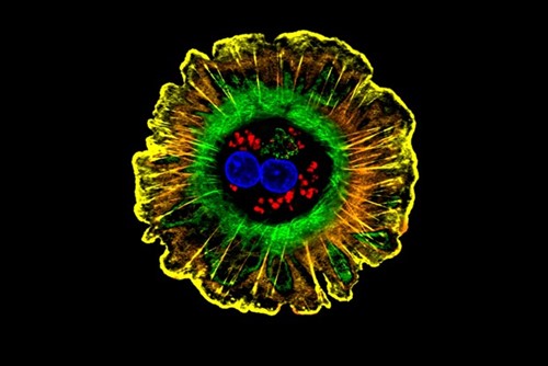
Image Credit: NIH
Tissue Model Reveals Key Players in Liver Regeneration
Anne Trafton | MIT News
Office
The human liver has amazing regeneration capabilities: Even if up to 70 percent of it is removed, the remaining tissue can regrow a full-sized liver within months.
Taking advantage of this regenerative capability could give doctors many more options for treating chronic liver disease. MIT engineers have now taken a step toward that goal, by creating a new liver tissue model that allows them to trace the steps involved in liver regeneration more precisely than has been possible before.
The new model can yield information that couldn’t be gleaned from studies of mice or other animals, whose biology is not identical to that of humans, says Sangeeta Bhatia, the leader of the research team.
“For years, people have been identifying different genes that seem to be involved in mouse liver regeneration, and some of them seem to be important in humans, but they have never managed to figure out all of the cues to make human liver cells proliferate,” says Bhatia, the John and Dorothy Wilson Professor of Health Sciences and Technology and of Electrical Engineering and Computer Science at MIT and a member of MIT’s Koch Institute for Integrative Cancer Research and Institute for Medical Engineering and Science.
The new study, which appears this week in the Proceedings of the National Academy of Sciences, has identified one molecule that appears to play a key role, and also yielded several other candidates that the researchers plan to explore further.
The lead author of the paper is Arnav Chhabra, a former MIT graduate student and postdoc.
Regeneration on a Chip
Most of the patients who need liver transplants suffer from chronic illnesses such as viral hepatitis, fatty liver disease, or cancer. However, if researchers had a reliable way to stimulate the liver to regenerate on its own, some transplants could be avoided, Bhatia says. Or, such stimulation might be used to help a donated liver grow after being transplanted.
From studies in mice, researchers have learned a great deal about some of the regeneration pathways that are activated after liver injury or illness. One key factor is the reciprocal relationship between hepatocytes (the main type of cell found in the liver) and endothelial cells, which line the blood vessels. Hepatocytes produce factors that help blood vessels develop, and endothelial cells generate growth factors that help hepatocytes proliferate.
Another contributor that researchers have identified is fluid flow in the blood vessels. In mice, an increase in blood flow can stimulate the endothelial cells to produce signals that promote regeneration.
To model all of these interactions, Bhatia’s lab teamed up with Christopher Chen, the William F. Warren Distinguished Professor of Biomedical Engineering at Boston University, who designs microfluidic devices with channels that mimic blood vessels. To create these models of “regeneration on a chip,” the researchers grew blood vessels along one of these microfluidic channels and then added multicellular spheroid aggregates derived from liver cells from human organ donors.
The chip is designed so that molecules such as growth factors can flow between the blood vessels and the liver spheroids. This setup also allows the researchers to easily knock out genes of interest in a specific cell type and then see how it affects the overall system.
Using this system, the researchers showed that increased fluid flow on its own did not stimulate hepatocytes to enter the cell division cycle. However, if they also delivered an inflammatory signal (the cytokine IL-1-beta), hepatocytes did enter the cell cycle.
When that happened, the researchers were able to measure what other factors were being produced. Some were expected based on earlier mouse studies, but others had not been seen before in human cells, including a molecule called prostaglandin E2 (PGE2).
The MIT team found high levels of this molecule, which is also involved in zebrafish regeneration, in their liver regeneration system. By knocking out the gene for PGE2 biosynthesis in endothelial cells, the researchers were able to show that those cells are the source of PGE2, and they also demonstrated that this molecule stimulates human liver cells to enter the cell cycle.
Human-Specific Pathways
The researchers now plan to further explore some of the other growth factors and molecules that are produced on their chip during liver regeneration.
“We can look at the proteins that are being produced and ask, what else on this list has the same pattern as the other molecules that stimulate cell division, but is novel?” Bhatia says. “We think we can use this to discover new human-specific pathways.”
In this study, the researchers focused on molecules that stimulate cells to enter cell division, but they now hope to follow the process further along and identify molecules needed to complete the cell cycle. They also hope to discover the signals that tell the liver when to stop regenerating.
Bhatia hopes that eventually researchers will be able to harness these molecules to help treat patients with liver failure. Another possibility is that doctors could use such factors as biomarkers to determine how likely it is that a patient’s liver will regrow on its own.
“Right now when patients come in with liver failure, you have to transplant them because you don’t know if they’re going to recover on their own. But if we knew who had a robust regenerative response, and if we just needed to stabilize them for a little while, we could spare those patients from transplant,” Bhatia says.
The research was funded in part by the National Institutes of Health, the National Science Foundation Graduate Research Fellowship Program, Wellcome Leap, and the Paul and Daisy Soros Fellowship Program.
Reprinted with the permission MIT News http://news.mit.edu/
|
Virtual Roadshow – August 23 Lineage Cell Therapeutics – Brian M. Culley, CEOJoin Lineage Cell Therapeutics CEO Brian M. Culley for this exclusive corporate presentation, followed by a Q & A session moderated by Robert LeBoyer, Noble’s senior research analyst, featuring questions taken from the audience. Registration is free and open to all investors, at any level Register Now
|
Suggested Content
 The Gene Therapy Space May be Blossoming
|
 Covid-19 Vaccine and Healthcare Responsibility to Begin Falling on the Patient
|
 Monkeypox Treatments and Vaccines are Creating Investor Opportunities
|
 Is Biotech’s Outperformance Reaching a New Stage of Development?
|
Stay up to date. Follow us:

|